Heavy exudate can unravel even the best care plan: leaking through bandages, soaking clothes and linens, and leaving the periwound skin pale, boggy, and fragile. This guide translates the evidence and day‑to‑day practice into a clear approach for choosing and using a super absorbent wound dressing to contain fluid, protect skin, and support the rest of the treatment plan. It is written for US clinicians and procurement teams managing venous leg ulcers under compression, diabetic foot ulcers with fluctuating output, postoperative incisions or drain sites, and stage III–IV pressure injuries.
What a Super Absorbent Wound Dressing Is and Why Retention Matters
A super absorbent wound dressing typically uses a multilayer construction with a distribution layer over a superabsorbent polymer core that sequesters exudate. Two concepts drive real‑world performance: absorption—how much fluid a dressing can take up; and retention or lock‑in—how securely that fluid is held when the dressing is pressed, bent, or loaded under bandaging. Under compression, retention becomes decisive. Laboratory methods described in the EN 13726 standard include assessments of absorption under compression and retention capacity, offering a shared language for comparing products, even if bench metrics do not perfectly predict bedside outcomes. For an overview of these methods, see the EN 13726 update explainer from NAMSA in 2024, which outlines how “absorption under compression” is tested in vitro and why it matters for clinical selection according to the EN 13726 update explainer by NAMSA (2024): EN 13726 updates on assessing wound dressing performance.
Assessing exudate and aiming for moisture balance
Most wound frameworks place moisture balance at the center of care. TIME and its extension TIMERS emphasize matching dressing capacity to output, protecting periwound skin, and reassessing frequently as bioburden and tissue status change. Practical assessment starts with how saturated the previous dressing was at removal, the presence of strike‑through, the look and feel of the wound surface, and signs of periwound maceration.
Below is a quick operational guide clinicians can adapt in documentation.
Exudate level | Observable cues at change | Typical dressing response target |
|---|---|---|
Low | Contact layer slightly damp; no strike‑through; wound surface moist | Light absorbers; extend wear time if safe |
Moderate | 25–75% pad saturation; edges moist; occasional weeping | Medium to high capacity absorbers; protect periwound |
Heavy | Pad near full saturation or strike‑through; pooling; periwound whitening or bogginess | High capacity super absorbents with strong lock‑in; change before saturation; barrier film |
According to a 2024 systematic review that synthesized mixed study designs, superabsorbent dressings are designed to sequester exudate and have been associated in observational settings with fewer leakage events and extended change intervals, though heterogeneity and funding sources warrant cautious interpretation. See the peer‑reviewed synthesis for details: Systematic review of superabsorbent dressings.
Selection at a glance for heavy exudate
Think of selection as pairing capacity with context: choose a super absorbent wound dressing when heavy or persistent exudate risks strike‑through or periwound maceration, or when output spikes under compression or device fixation. Decide the wound contact interface based on skin status and fixation needs—a non‑adherent contact layer when you plan to secure with secondary fixation, or a soft silicone contact layer when atraumatic removal and gentle adhesion are priorities. Under compression, prefer thinner profiles that will not distort sub‑bandage pressure, and monitor interface pressure when introducing thicker absorbers.
Venous leg ulcers under compression
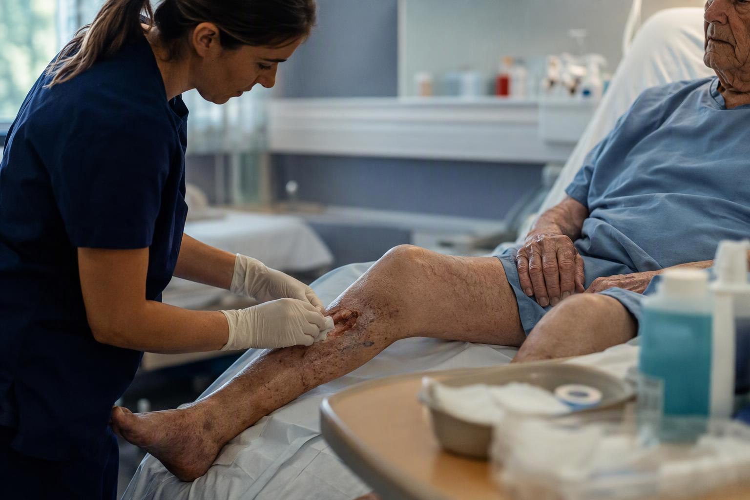
Compression remains foundational therapy for venous leg ulcers. The dressing’s job is to contain fluid and protect the surrounding skin while not compromising the pressure system. Match capacity to output and prioritize retention under pressure to help limit strike‑through beneath multilayer wraps. Size to extend beyond the wound by at least 1–2 cm without bunching. Protect the periwound with an alcohol‑free barrier film when the skin looks pale, sodden, or fragile. Start with closer reassessment intervals when output is copious, then extend wear time as moisture control stabilizes. A neutral workflow example: If the periwound is fragile and the leg will be rewrapped frequently, a silicone‑adherent superabsorber can provide gentle fixation and atraumatic removal beneath compression. For a product specification example, see the silicone‑adherent super absorbent dressing page at SLK Medical.
Evidence notes for clinicians and procurement: Economic modeling in 2022 suggested potential cost‑in‑use advantages of superabsorbents over foams for moderate to high exudate in venous leg ulcers, largely via fewer changes and reduced leakage‑related resource use. Review the model details here: Cost‑effectiveness analysis for venous leg ulcers. Case series and evaluations report practical success using superabsorbents under multilayer compression; however, large comparative trials with maceration endpoints are limited. Use your local outcomes tracking to validate change intervals and leakage rates.
Diabetic foot ulcers with fluctuating output and infection risk
Two pillars come first for plantar ulcers: offloading and infection control. Dressings support the plan by managing moisture and protecting periwound skin. For non‑infected wounds, current guidance from the International Working Group on the Diabetic Foot advises against routine antimicrobial or antiseptic dressings to promote healing. See the 2023 recommendation: IWGDF Wound Healing Guideline 2023. When heavy exudate threatens maceration, select a super absorbent wound dressing and set the change interval to stay ahead of saturation. A silicone contact layer can reduce trauma at the plantar margin during frequent checks. If local signs suggest critical colonization or superficial infection, follow institutional protocols and infection guidance from the IDSA/IWGDF while addressing systemic needs; avoid assuming that antimicrobial dressings alone will resolve infection.
Postoperative incisions and drain sites
In the first days after surgery, some incisions and drain sites produce moderate to high serous output or lymphorrhea. Here, moisture control coexists with the principle of undisturbed wound healing whenever feasible. For copious output, select high‑capacity absorbency and secure fixation while avoiding skin trauma at the edges. Superabsorbent pads can serve as the primary absorber with fixation chosen to match skin integrity. Reassess early to calibrate change frequency based on saturation rather than fixed schedules. If output tapers quickly, transition to a lower‑capacity dressing and extend wear. Maintain aseptic handling and consider negative pressure wound therapy when output is persistent and high despite appropriate absorbers, per local protocols. For a concise overview of dressing classes and their use in surgical contexts, see the clinical summary in StatPearls: Wound dressings overview.
Stage III–IV pressure injuries and periwound protection
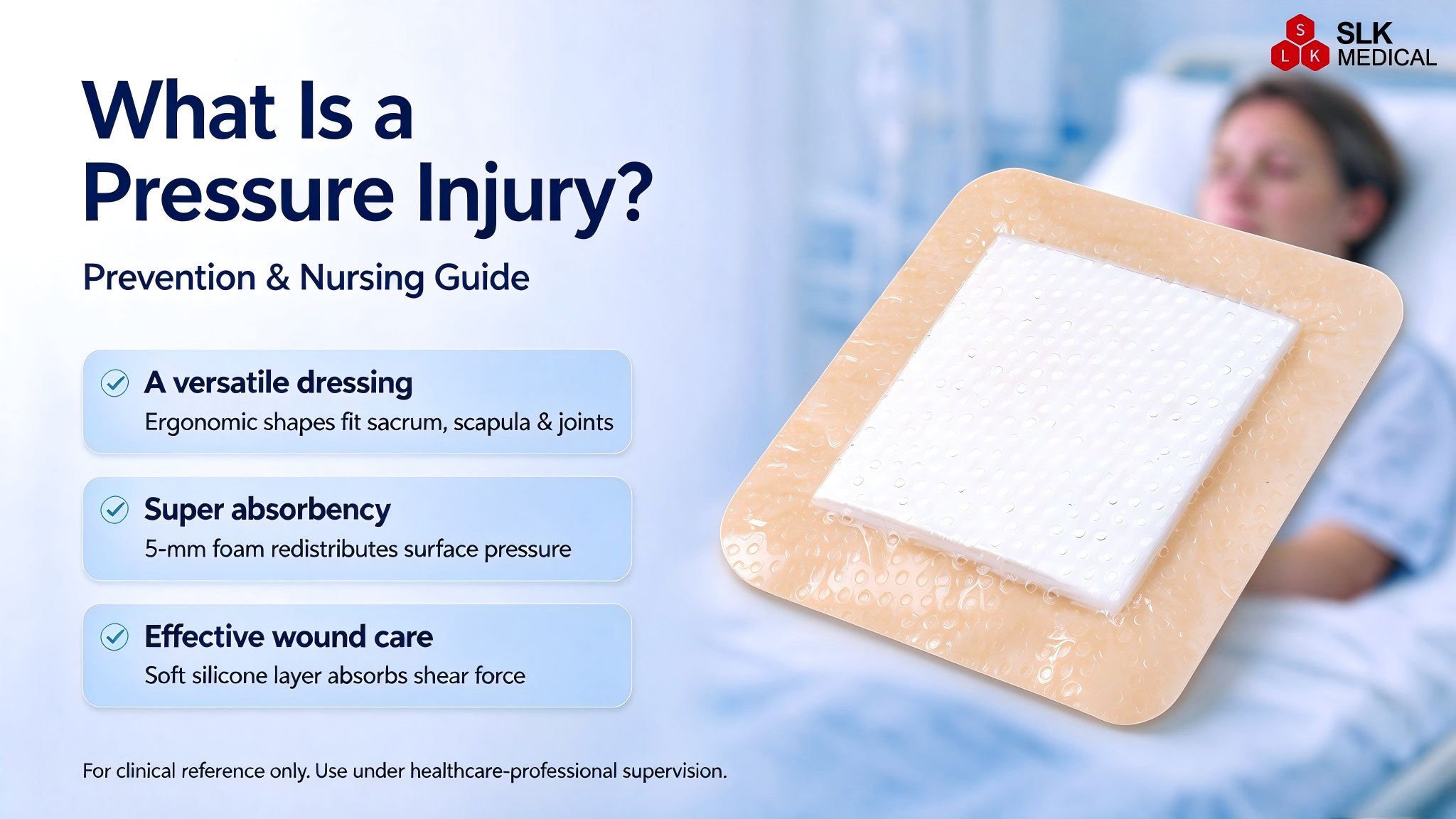
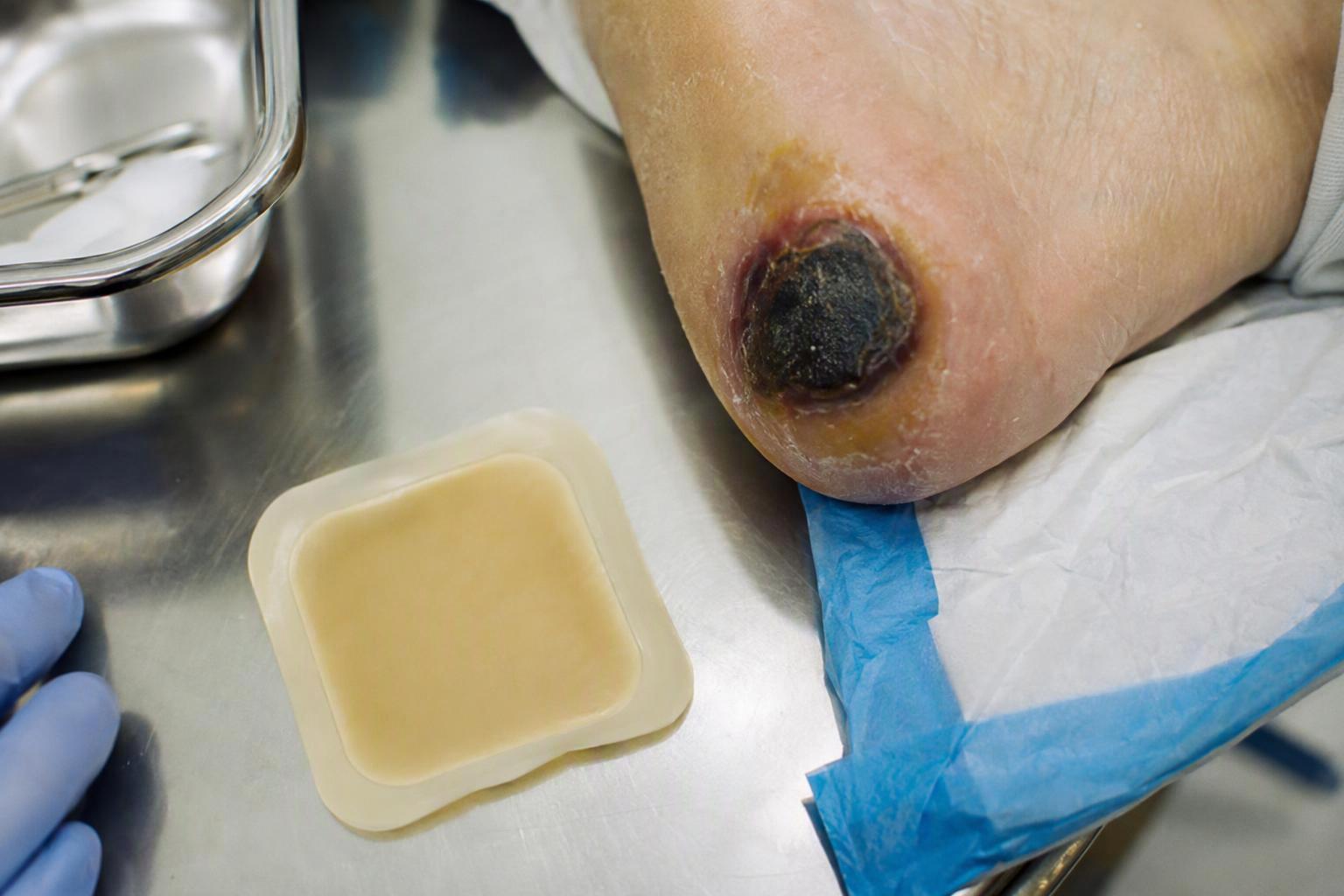
For full‑thickness pressure injuries, moisture control is part of a broader plan that includes pressure redistribution, nutrition, and bioburden management. Use a super absorbent wound dressing when exudate is heavy or persistent and threatens the periwound. A silicone contact layer can reduce skin stripping during frequent changes. Protect adjacent skin with barrier films or creams, especially where adhesive borders contact fragile tissue. International guidance hubs from NPIAP, EPUAP, and PPPIA emphasize moisture balance and periwound protection principles in their treatment chapters: International guideline information hubs. Consider negative pressure wound therapy for deep cavities or undermining when appropriate and available.
How superabsorbent compares with other absorbers
Clinicians often weigh superabsorbents against foams, alginates, and hydrofiber dressings. The table below provides a quick, non‑brand‑specific comparison to orient selection. Always confirm specific product capabilities from manufacturer instructions for use and your formulary notes.
Dressing class | Absorption capacity | Retention under pressure | Typical wear time guidance | Common indications in heavy exudate |
|---|---|---|---|---|
Superabsorbent core pad | Very high | High with strong lock‑in | Change before saturation; may extend as control improves | VLUs under compression, DFUs with maceration risk, high‑output surgical sites, Stage III–IV pressure injuries |
Foam (incl. silicone border) | Moderate to high | Moderate; varies by brand and thickness | 2–4 days typical in moderate output | Moderate exudate wounds; prophylaxis and comfort where capacity suffices |
Alginate | High initial uptake | Lower lock‑in; gels with fluid | Change based on gel saturation and strike‑through | Cavities and irregular shapes; often combined with secondary absorbers |
Hydrofiber | High uptake with gel formation | Variable; depends on secondary fixation | Based on saturation; avoid overdrying | Cavities and undermining; conforming filler with secondary absorbers |
A 2024 peer‑reviewed synthesis noted observational trends toward fewer leakage events and extended intervals with superabsorbents in exudate‑heavy wounds, though study heterogeneity limits firm comparative conclusions. For details, see the 2024 systematic review of superabsorbent dressings. For testing context and why bench retention matters, review the EN 13726 updates on absorption under compression.
Preventing periwound maceration
If you could only do three things to prevent periwound breakdown while using a super absorbent wound dressing, what would they be? Here’s a pragmatic toolkit grounded in evidence and everyday practice: set change frequency by saturation (start 24–48 hours in heavy output, then extend), shield the periwound with an alcohol‑free barrier film when skin looks blanchy or boggy, and choose a skin‑friendly interface such as a silicone contact layer to reduce trauma during frequent checks. Educational resources and clinical summaries describe reduced maceration when films are used consistently beneath adhesives and around high‑output wounds; a helpful overview appears in the Wounds UK quick guide to maceration management: Quick guide to periwound maceration. For a plain‑language explainer on superabsorbent mechanisms and clinical use, see the clinician‑facing article at WoundSource: What are superabsorbent dressings.
Stepwise application and removal protocol
A super absorbent wound dressing works best when the fundamentals are consistent. The following sequence can be adapted to your protocol and the care setting.
Prepare the site: Cleanse per policy, gently dry the periwound, and apply an alcohol‑free barrier film where skin is at risk. Debride and manage bioburden per your institutional protocol.
Select size and interface: Choose a pad that extends 1–2 cm beyond the wound edge. Use a silicone contact layer when atraumatic removal is a priority or when adhesives frequently contact fragile skin.
Apply and secure: Lay the dressing flat without creases. Use appropriate fixation—retention bandage, secondary cover, or silicone border—ensuring the profile will not create pressure points under compression.
Document and reassess: Record exudate level, pad saturation, periwound status, and any leakage or odor. For copious output, reassess within 24–48 hours, then extend as control improves.
Remove gently: Lift silicone interfaces slowly at a low angle; inspect the periwound for whitening, fragility, or denudation. Reapply barrier film as needed before the next dressing.
Clinical nuance under compression: When you add or change absorber thickness in a compression system, recheck sub‑bandage pressures if you have access to interface pressure tools, and watch for pressure points over bony prominences.
Troubleshooting and escalation guide
Persistent strike‑through despite timely changes: Increase capacity, improve seal and fixation, reassess compression technique, and consider negative pressure wound therapy if appropriate.
Periwound whitening or denudation: Shorten the change interval, intensify barrier film use, and evaluate whether fluid is laterally wicking to the edges.
Sudden increase in output with odor or pain: Reassess for infection; obtain cultures as indicated; align with antimicrobial stewardship and systemic therapy per IDSA/IWGDF guidance.
Pain on removal or skin stripping: Switch to or add a silicone contact layer; use adhesive remover if compatible with your products and policies.
For international guidance anchors on pressure injury treatment principles and periwound protection, consult the NPIAP/EPUAP/PPPIA guideline information hubs.
Procurement and documentation checklist
For formulary decisions and charting, keep these points front and center: sizes and formats that fit your common wound locations and compression protocols; interface types available for fragile skin and frequent rechecks; sterility and packaging that aligns with your unit’s throughput and storage; instructions for absorption under compression and retention data where available; and documentation fields capturing exudate level, saturation at change, periwound status, and dressing change interval rationale.
References you can cite with patients and teams
Laboratory testing context for absorption and retention under load: EN 13726 updates on assessing wound dressing performance
DFU antimicrobial stance for non‑infected ulcers: IWGDF Wound Healing Guideline 2023
Pressure injury care principles and periwound protection: International guideline information hubs
Clinical overview of dressing classes for surgical contexts: StatPearls Wound Dressings
Synthesis of clinical evidence on superabsorbents: 2024 systematic review of superabsorbent dressings
Economic model for venous leg ulcers: Cost‑effectiveness analysis for venous leg ulcers
Next steps
If you are aligning protocols or building a formulary for heavy exudate, review specifications and size availability across formats. For a consolidated view of options, browse the super absorbent category archive at SLK Medical — Super Absorbent Dressing category.
Notes on scope and safety: This guide is instructional and does not replace clinical judgment. Always follow institutional policies, manufacturer instructions for use, and patient‑specific considerations, including compression therapy for venous disease and offloading for plantar diabetic ulcers.